MIND Technology Platform
Revolutionary integration of ultra-high field MRI and lightsheet microscopy
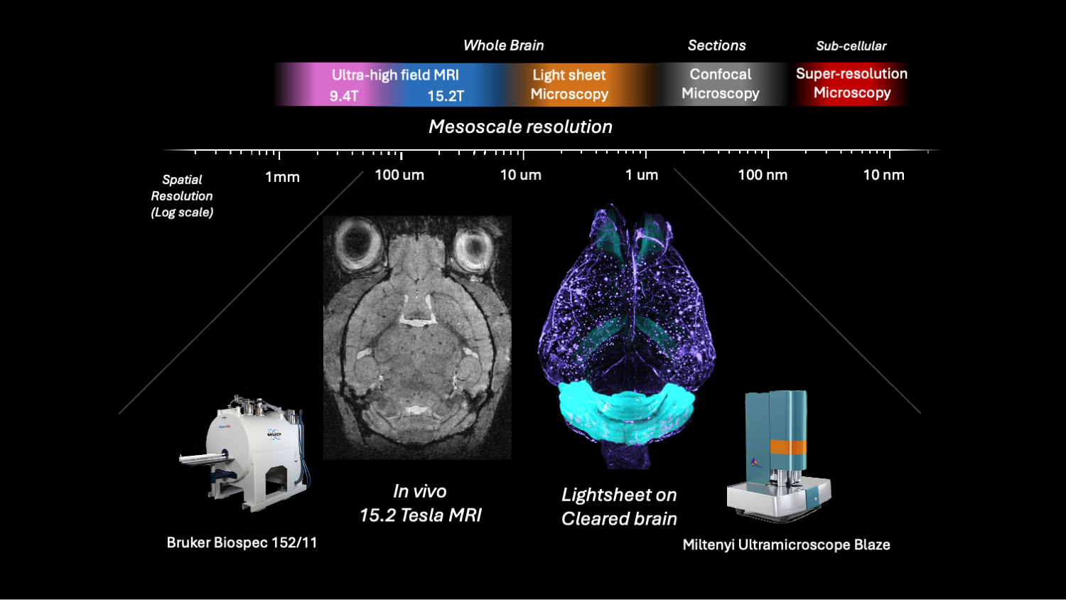
Integration of MRI (macro-scale) and lightsheet microscopy (micro-scale) for comprehensive brain analysis
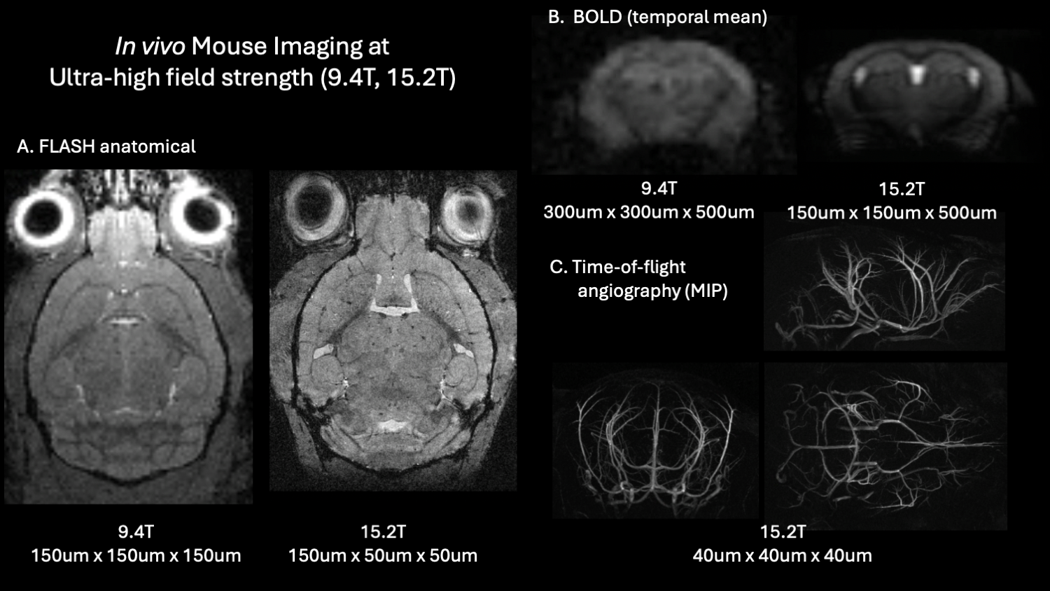
Enhanced resolution and contrast at 15.2T enables detection of subtle brain changes
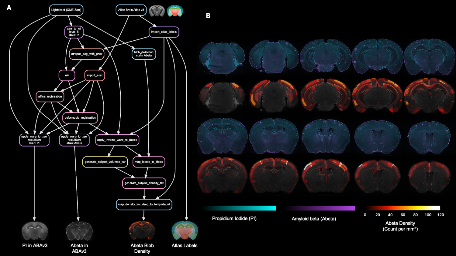
Automated pipeline from raw lightsheet data to quantitative pathology maps
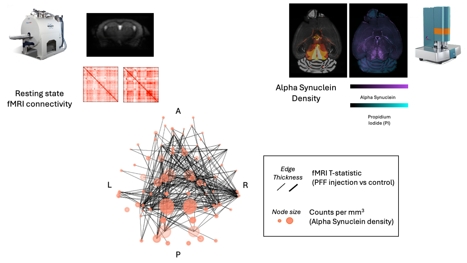
Integration of resting-state fMRI networks with alpha-synuclein pathology mapping
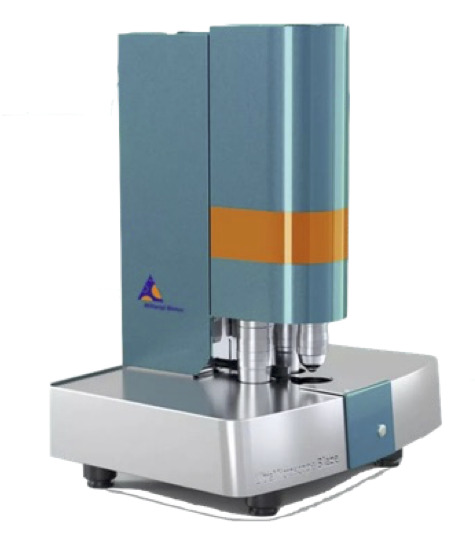
Miltenyi Blaze Ultramicroscope 2
Advanced lightsheet microscopy system featuring dual-side illumination, multi-channel fluorescence detection, and automated sample handling for high-throughput whole-brain imaging.
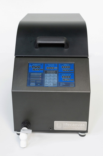
LifeCanvas SmartBatch Clearing
Automated tissue clearing system enabling batch processing of brain samples with optimized protocols for enhanced transparency and preserved fluorescence.
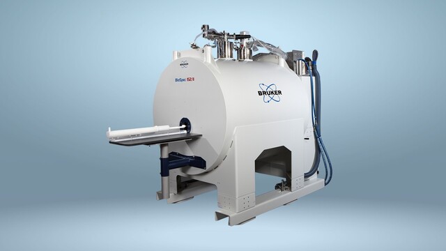
Bruker 15.2T MRI Scanner
Ultra-high field MRI system providing exceptional spatial resolution and contrast sensitivity for advanced neuroimaging applications and connectivity mapping.